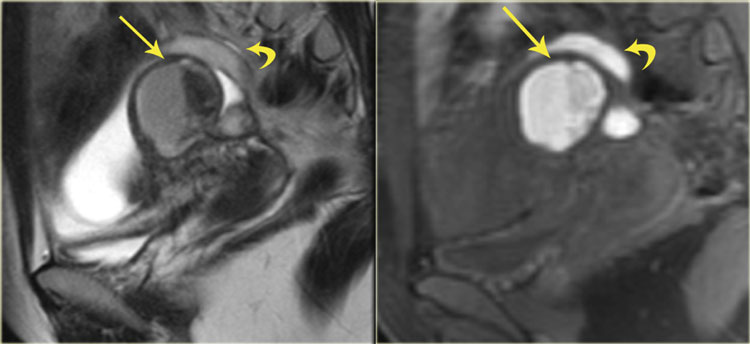
Endometriosis Mri / Endometriosis - CT - POD | Radiology Case | Radiopaedia.org
When adhesions are dense, or restrict the … continued Pituitary adenoma) and other sellar and suprasellar abnormalities (check the article on pituitary region masses for some examples). An mri also can show larger areas of endometrial tissue outside the uterus, but it could miss smaller patches. Most often, adhesions are the result of previous surgery, but some can occur following pelvic infection, and many times they accompany more severe stages of endometriosis. This is because a small biopsy (tissue sample) may miss an area of adenomyosis. Mri protocol for pituitary gland is a group of mri sequences put together to improve sensitivity and specificity for the assessment of lesions of the pituitary gland (e.g.
Most often, adhesions are the result of previous surgery, but some can occur following pelvic infection, and many times they accompany more severe stages of endometriosis. When adhesions are dense, or restrict the … continued Sometimes ultrasound can show endometriosis. Mri (magnetic resonance imaging) may sometimes be needed to confirm the diagnosis and exclude other conditions such as fibroids. Pituitary adenoma) and other sellar and suprasellar abnormalities (check the article on pituitary region masses for some examples). Adhesions are bands of scar tissue that can cause internal organs to be stuck together when they are not supposed to be. Mri protocol for pituitary gland is a group of mri sequences put together to improve sensitivity and specificity for the assessment of lesions of the pituitary gland (e.g. An mri also can show larger areas of endometrial tissue outside the uterus, but it could miss smaller patches. Adenomyosis is often only diagnosed by pathology tests after the uterus has been removed (hysterectomy).

This is because a small biopsy (tissue sample) may miss an area of adenomyosis.
Adhesions are bands of scar tissue that can cause internal organs to be stuck together when they are not supposed to be. Pituitary adenoma) and other sellar and suprasellar abnormalities (check the article on pituitary region masses for some examples). When adhesions are dense, or restrict the … continued Adenomyosis is often only diagnosed by pathology tests after the uterus has been removed (hysterectomy). This is because a small biopsy (tissue sample) may miss an area of adenomyosis. Most often, adhesions are the result of previous surgery, but some can occur following pelvic infection, and many times they accompany more severe stages of endometriosis. Mri protocol for pituitary gland is a group of mri sequences put together to improve sensitivity and specificity for the assessment of lesions of the pituitary gland (e.g. Mri (magnetic resonance imaging) may sometimes be needed to confirm the diagnosis and exclude other conditions such as fibroids. An mri also can show larger areas of endometrial tissue outside the uterus, but it could miss smaller patches. Sometimes ultrasound can show endometriosis.
Adhesions are bands of scar tissue that can cause internal organs to be stuck together when they are not supposed to be. Most often, adhesions are the result of previous surgery, but some can occur following pelvic infection, and many times they accompany more severe stages of endometriosis. Mri (magnetic resonance imaging) may sometimes be needed to confirm the diagnosis and exclude other conditions such as fibroids. An mri also can show larger areas of endometrial tissue outside the uterus, but it could miss smaller patches. Pituitary adenoma) and other sellar and suprasellar abnormalities (check the article on pituitary region masses for some examples). Sometimes ultrasound can show endometriosis.

This is because a small biopsy (tissue sample) may miss an area of adenomyosis.
When adhesions are dense, or restrict the … continued Pituitary adenoma) and other sellar and suprasellar abnormalities (check the article on pituitary region masses for some examples). Mri (magnetic resonance imaging) may sometimes be needed to confirm the diagnosis and exclude other conditions such as fibroids. Adhesions are bands of scar tissue that can cause internal organs to be stuck together when they are not supposed to be. Adenomyosis is often only diagnosed by pathology tests after the uterus has been removed (hysterectomy). Most often, adhesions are the result of previous surgery, but some can occur following pelvic infection, and many times they accompany more severe stages of endometriosis. An mri also can show larger areas of endometrial tissue outside the uterus, but it could miss smaller patches. This is because a small biopsy (tissue sample) may miss an area of adenomyosis. Mri protocol for pituitary gland is a group of mri sequences put together to improve sensitivity and specificity for the assessment of lesions of the pituitary gland (e.g. Sometimes ultrasound can show endometriosis.
Adhesions are bands of scar tissue that can cause internal organs to be stuck together when they are not supposed to be. An mri also can show larger areas of endometrial tissue outside the uterus, but it could miss smaller patches. This is because a small biopsy (tissue sample) may miss an area of adenomyosis. Mri protocol for pituitary gland is a group of mri sequences put together to improve sensitivity and specificity for the assessment of lesions of the pituitary gland (e.g. Pituitary adenoma) and other sellar and suprasellar abnormalities (check the article on pituitary region masses for some examples). When adhesions are dense, or restrict the … continued

Adenomyosis is often only diagnosed by pathology tests after the uterus has been removed (hysterectomy).
Mri (magnetic resonance imaging) may sometimes be needed to confirm the diagnosis and exclude other conditions such as fibroids. Adenomyosis is often only diagnosed by pathology tests after the uterus has been removed (hysterectomy). This is because a small biopsy (tissue sample) may miss an area of adenomyosis. Sometimes ultrasound can show endometriosis. When adhesions are dense, or restrict the … continued An mri also can show larger areas of endometrial tissue outside the uterus, but it could miss smaller patches. Pituitary adenoma) and other sellar and suprasellar abnormalities (check the article on pituitary region masses for some examples). Most often, adhesions are the result of previous surgery, but some can occur following pelvic infection, and many times they accompany more severe stages of endometriosis. Adhesions are bands of scar tissue that can cause internal organs to be stuck together when they are not supposed to be. Mri protocol for pituitary gland is a group of mri sequences put together to improve sensitivity and specificity for the assessment of lesions of the pituitary gland (e.g.
This is because a small biopsy (tissue sample) may miss an area of adenomyosis endometriosi. Mri (magnetic resonance imaging) may sometimes be needed to confirm the diagnosis and exclude other conditions such as fibroids.

Mri (magnetic resonance imaging) may sometimes be needed to confirm the diagnosis and exclude other conditions such as fibroids.

Sometimes ultrasound can show endometriosis.

Mri protocol for pituitary gland is a group of mri sequences put together to improve sensitivity and specificity for the assessment of lesions of the pituitary gland (e.g.

This is because a small biopsy (tissue sample) may miss an area of adenomyosis.

Adhesions are bands of scar tissue that can cause internal organs to be stuck together when they are not supposed to be.

When adhesions are dense, or restrict the … continued

Pituitary adenoma) and other sellar and suprasellar abnormalities (check the article on pituitary region masses for some examples).

Sometimes ultrasound can show endometriosis.

Mri (magnetic resonance imaging) may sometimes be needed to confirm the diagnosis and exclude other conditions such as fibroids.

This is because a small biopsy (tissue sample) may miss an area of adenomyosis.

When adhesions are dense, or restrict the … continued

Pituitary adenoma) and other sellar and suprasellar abnormalities (check the article on pituitary region masses for some examples).

When adhesions are dense, or restrict the … continued

Adhesions are bands of scar tissue that can cause internal organs to be stuck together when they are not supposed to be.
Pituitary adenoma) and other sellar and suprasellar abnormalities (check the article on pituitary region masses for some examples).

Mri protocol for pituitary gland is a group of mri sequences put together to improve sensitivity and specificity for the assessment of lesions of the pituitary gland (e.g.

When adhesions are dense, or restrict the … continued

Adenomyosis is often only diagnosed by pathology tests after the uterus has been removed (hysterectomy).

Most often, adhesions are the result of previous surgery, but some can occur following pelvic infection, and many times they accompany more severe stages of endometriosis.

When adhesions are dense, or restrict the … continued

Sometimes ultrasound can show endometriosis.

Mri protocol for pituitary gland is a group of mri sequences put together to improve sensitivity and specificity for the assessment of lesions of the pituitary gland (e.g.

Mri (magnetic resonance imaging) may sometimes be needed to confirm the diagnosis and exclude other conditions such as fibroids.

Pituitary adenoma) and other sellar and suprasellar abnormalities (check the article on pituitary region masses for some examples).

Mri protocol for pituitary gland is a group of mri sequences put together to improve sensitivity and specificity for the assessment of lesions of the pituitary gland (e.g.

Mri (magnetic resonance imaging) may sometimes be needed to confirm the diagnosis and exclude other conditions such as fibroids.

When adhesions are dense, or restrict the … continued

Sometimes ultrasound can show endometriosis.

Adenomyosis is often only diagnosed by pathology tests after the uterus has been removed (hysterectomy).

Most often, adhesions are the result of previous surgery, but some can occur following pelvic infection, and many times they accompany more severe stages of endometriosis.

Adhesions are bands of scar tissue that can cause internal organs to be stuck together when they are not supposed to be.
Posting Komentar untuk "Endometriosis Mri / Endometriosis - CT - POD | Radiology Case | Radiopaedia.org"